
全部商品 / LEICA Light Sheet 層光掃描螢光影像系統 / LEICA Viventis 層光掃描顯微鏡系統

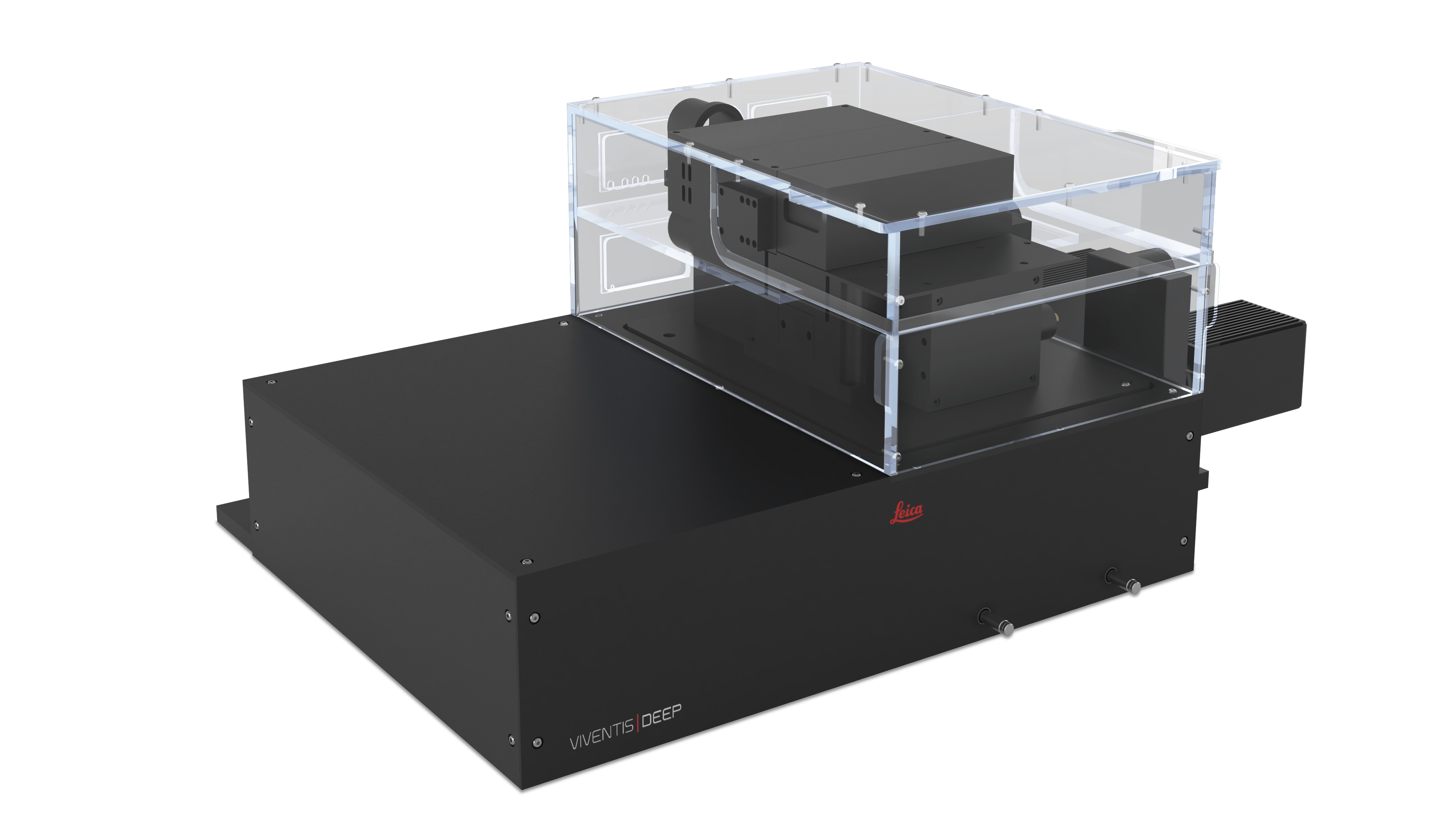
LEICA Viventis DEEP 雙側層光掃描顯微影像系統
LEICA 獨家創新的 Viventis DEEP 雙側層光掃描與雙側偵測成像顯微鏡系統 ( Dual View ), 唯一可同時提供
1). 多視角及多位置的 多樣本的同時成像 (Light Sheet 影像)
2). 高通量,
3). 超長時間,
4). 超高解析,
5). 光操控刺激模組
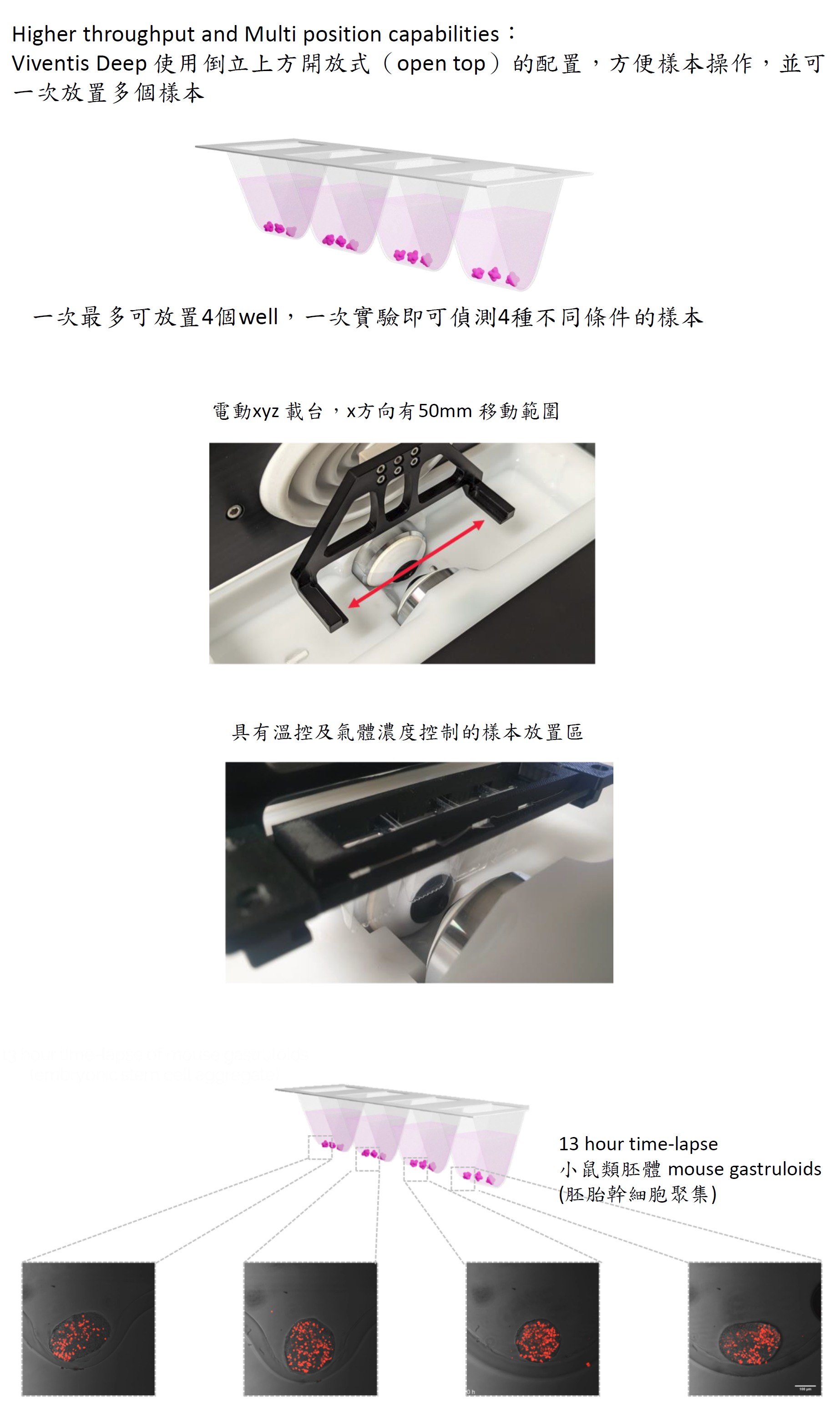
6). 上方開放式, 4-組樣本槽
7). 培養環境溫度與氣體濃度控制
觀察大型模式生物 (類器官) 深層細節及長時間動態變化。有效應用於大型生物器官, 類器官, 腸道類器官, 胚胎. 卵母細胞 .....等多模式生物樣本的 3D / 4D 成像長時間成像, 高通量, 多樣本的同時成像, 超高解析至長時間單一活細胞成像, 極低光毒性, 特別適合光敏感的活性樣本成像應用.
1). 多視角及多位置的 多樣本的同時成像 (Light Sheet 影像)
2). 高通量,
3). 超長時間,
4). 超高解析,
5). 光操控刺激模組
6). 上方開放式, 4-組樣本槽
7). 培養環境溫度與氣體濃度控制
觀察大型模式生物 (類器官) 深層細節及長時間動態變化。有效應用於大型生物器官, 類器官, 腸道類器官, 胚胎. 卵母細胞 .....等多模式生物樣本的 3D / 4D 成像長時間成像, 高通量, 多樣本的同時成像, 超高解析至長時間單一活細胞成像, 極低光毒性, 特別適合光敏感的活性樣本成像應用.
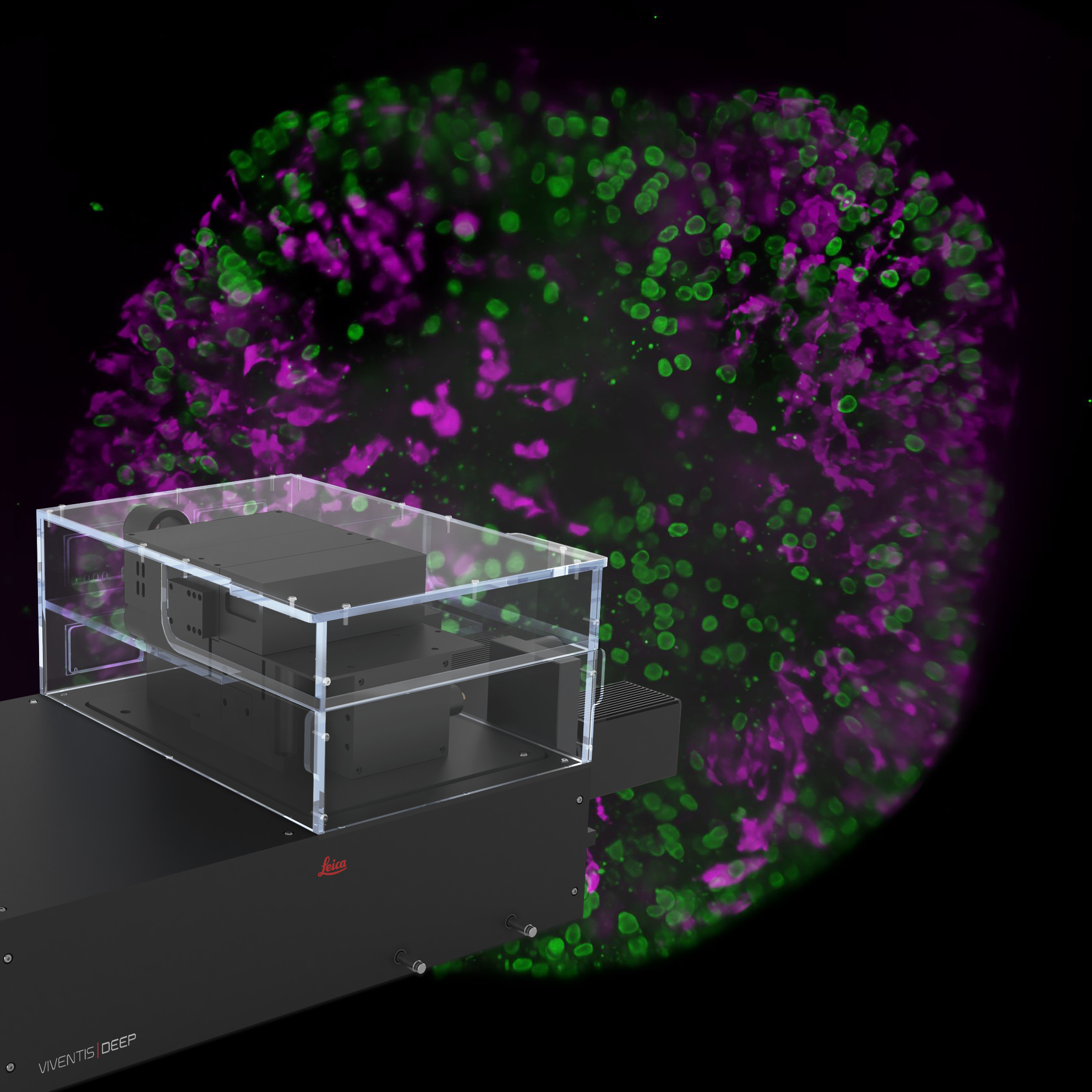
Viventis Deep 特點 :
雙側雷射層光掃描 與 偵測 :
解決了樣本深層光訊號散射的條紋偽影干擾問題, 結合了多視野 (multi-view) 和多位置 (multi-position) 的層光螢光成像, 實現樣本深層影像的超高解析, 一覽無遺 !
長時間成像, 極低光毒性
高通量, 多樣本的同時成像
應用於大型類器官, 同時也有效應用於活細胞成像
解決了樣本深層光訊號散射的條紋偽影干擾問題, 結合了多視野 (multi-view) 和多位置 (multi-position) 的層光螢光成像, 實現樣本深層影像的超高解析, 一覽無遺 !
長時間成像, 極低光毒性
高通量, 多樣本的同時成像
應用於大型類器官, 同時也有效應用於活細胞成像

Viventis Deep
以獨特方式提供多視角與多位置光片成像,全面呈現樣本的細節 。具有 高度空間和時間解析度 與影像品質,即使面對大型光散射樣本,仍能確保清晰成像 。
Viventis Deep 獨家技術
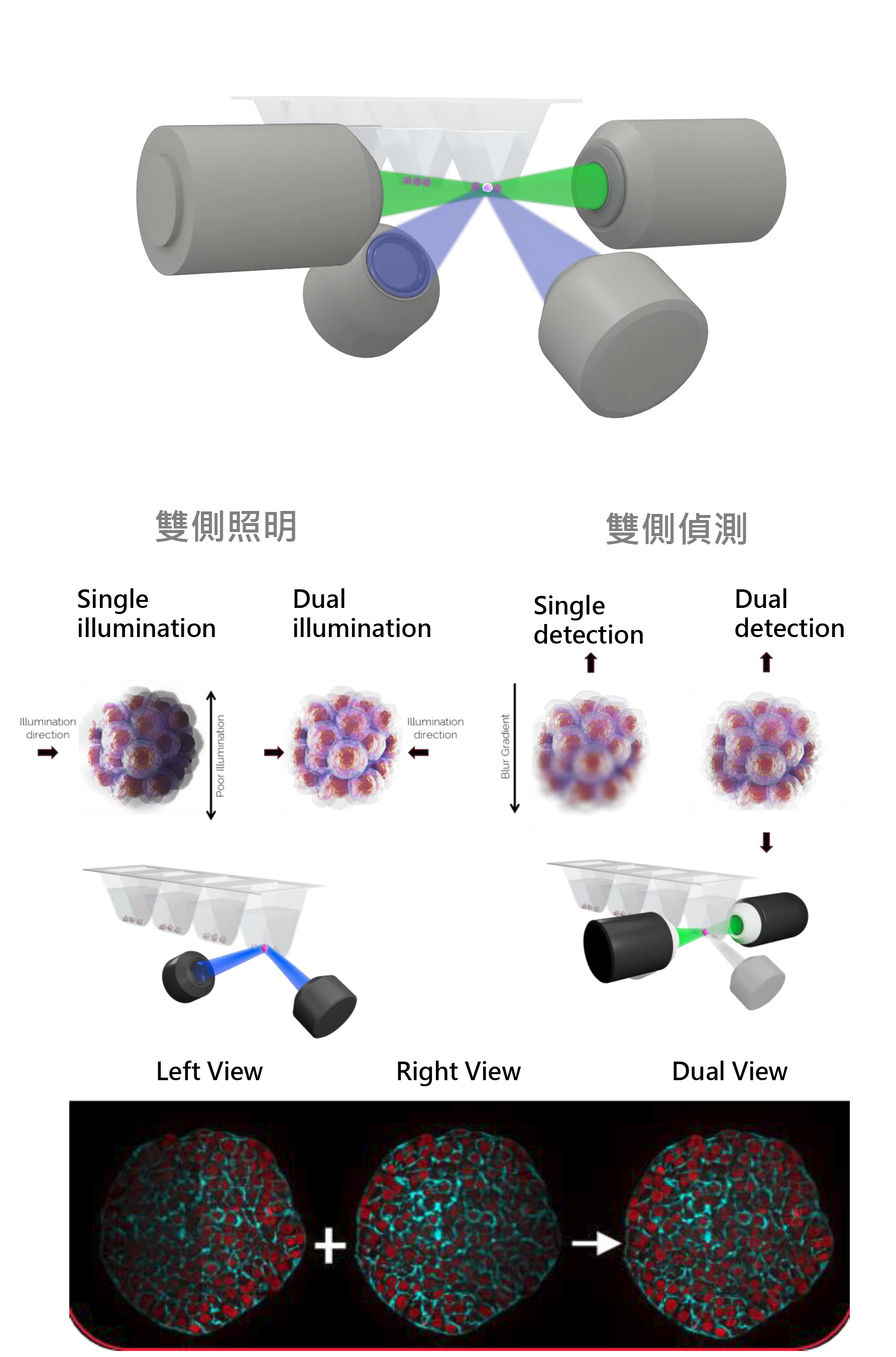
提供雙側照明及偵測以 " 解決樣本深層光訊號散射 " 的問題
提供雙側照明及偵測以 " 解決樣本深層光訊號散射 " 的問題
雙側照明樣本,而非單側照明,
可提升訊號強度並確保樣本整體訊號均勻分佈 。此外 ,雙側照明還能減少 light sheet 顯微技術固有的條紋偽影,提高成像品質
雙側偵測, 可同時從兩個方向成像樣本,
對於大型樣本尤為重要,有助於在 整個樣本 體積內清晰觀察單個細胞

Viventis Deep 使用雙側照明及偵測, 足以 " 解決樣本深層光訊號散射, 有效解決 light sheet 會造成條紋偽影的困擾 "
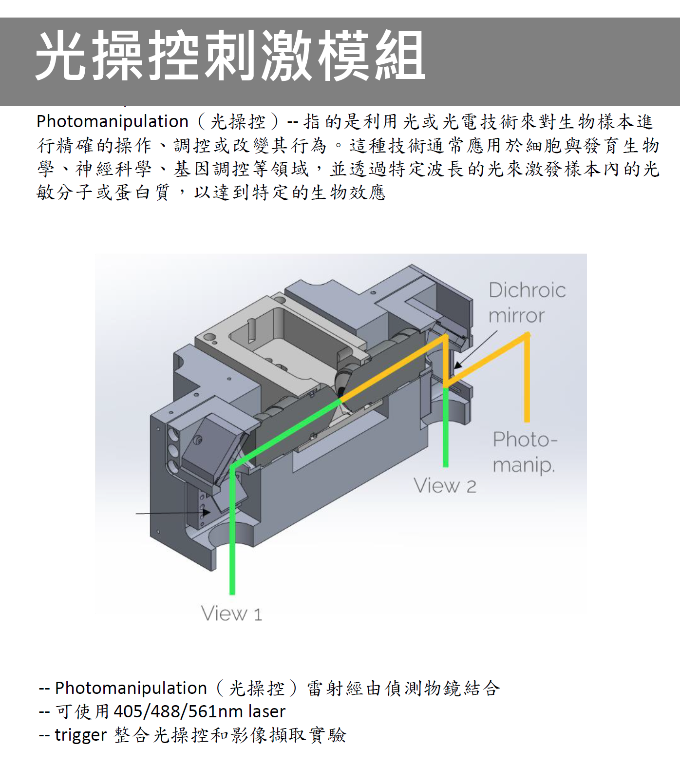

唯一可以同時提供
1). 多視角及多位置的 多樣本的同時成像 (Light Sheet 影像)
2). 高通量,
3). 超長時間,
4). 超高解析,
觀察大型模式生物 (類器官) 深層細節及長時間動態變化。有效應用於大型生物器官, 類器官, 腸道類器官, 胚胎. 卵母細胞 .....等多模式生物樣本的 3D / 4D 成像長時間成像, 高通量, 多樣本的同時成像, 超高解析至長時間單一活細胞成像, 極低光毒性, 特別適合光敏感的活性樣本成像應用.

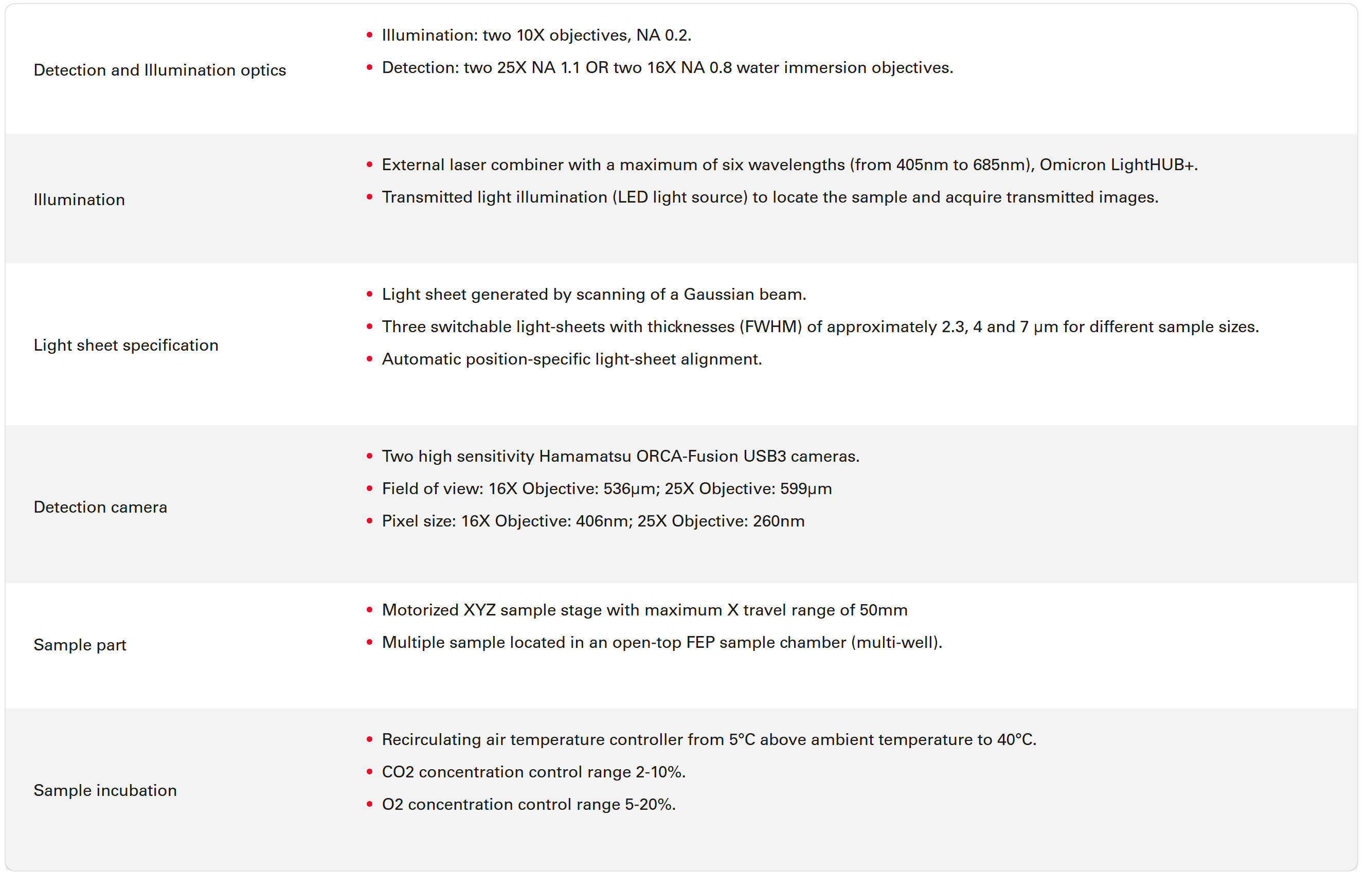

The Viventis Deep microscope helps you to expand the spatio-temporal understanding of your sample to its full depth, thanks to increased spatio-temporal resolution.
Achieve detailed volumetric imaging for a complete view of the sample with a patented combination of
- Dual illumination
- Dual view detection
- Multi-position
- Open top sample holder
You can even image large light scattering samples over time with outstanding quality for meaningful downstream analysis, while minimizing light dose and maintaining sample accessibility.
多細胞系統會在數周內從單個細胞生長成為類器官、由數千個細胞組成完整組織,此類樣品的即時成像一直具有挑戰性。為了跨越這些長時間、空間尺度的難題, Viventis Deep 採用開放式頂部雙側成像和雙側照明的光片顯微鏡,專門用於單細胞解析度的發育過程中的大型類器官樣本的即時成像。(如成熟的腸道類器官等) 中可以獲得定量單細胞資訊的能力。
腸道類器官長時間成像應用範例
Open-top multisample dual-view light-sheet microscope for live imaging of large multicellular systems | Nature Methods
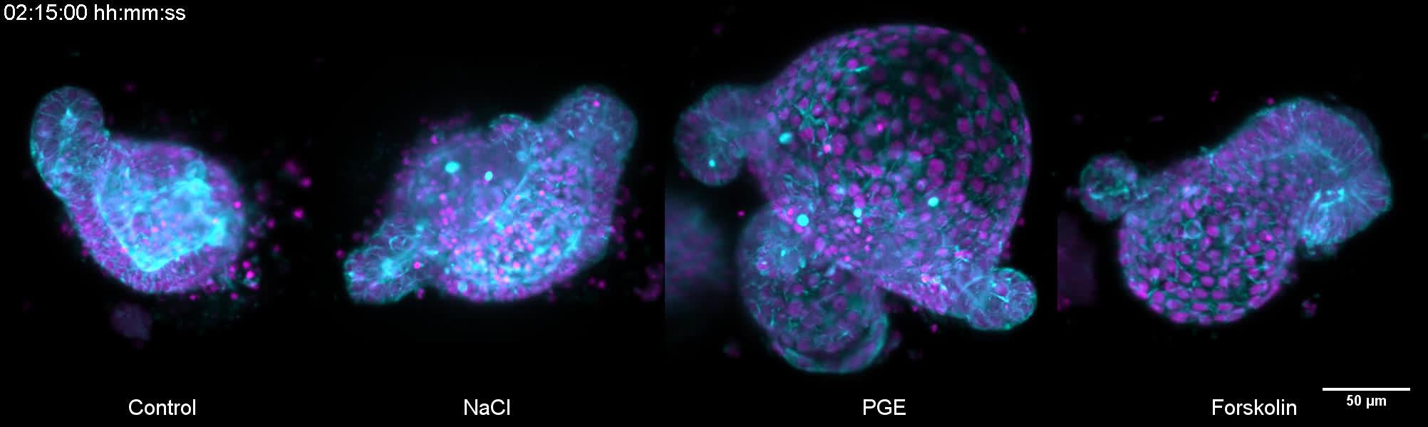
腸道類器官尺寸從200μm到550μm的多種樣品進行了成像,並進行了長達12天的連續成像:小鼠肝類器官,人類結腸癌類器官,小鼠腮腺唾液腺類器官,和類原腸胚。PGE、Forskolin和NaCl誘導的高滲休克對腸道類器官的機械滲透作用。
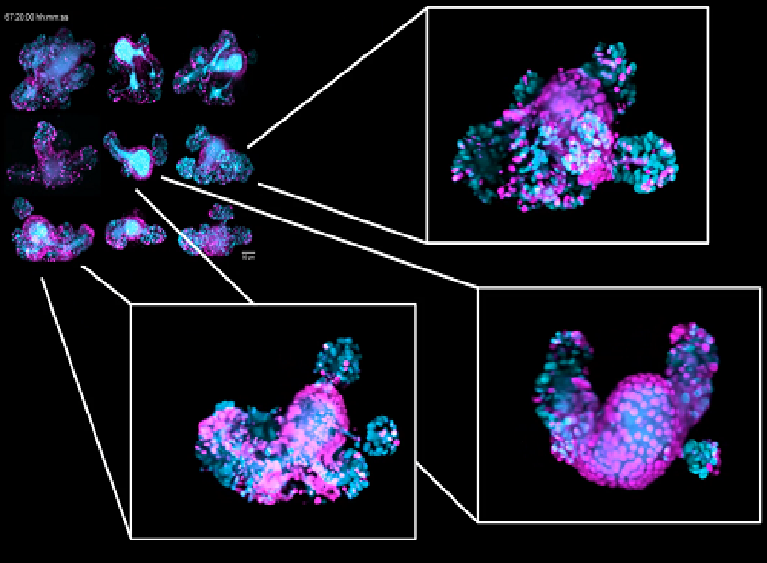
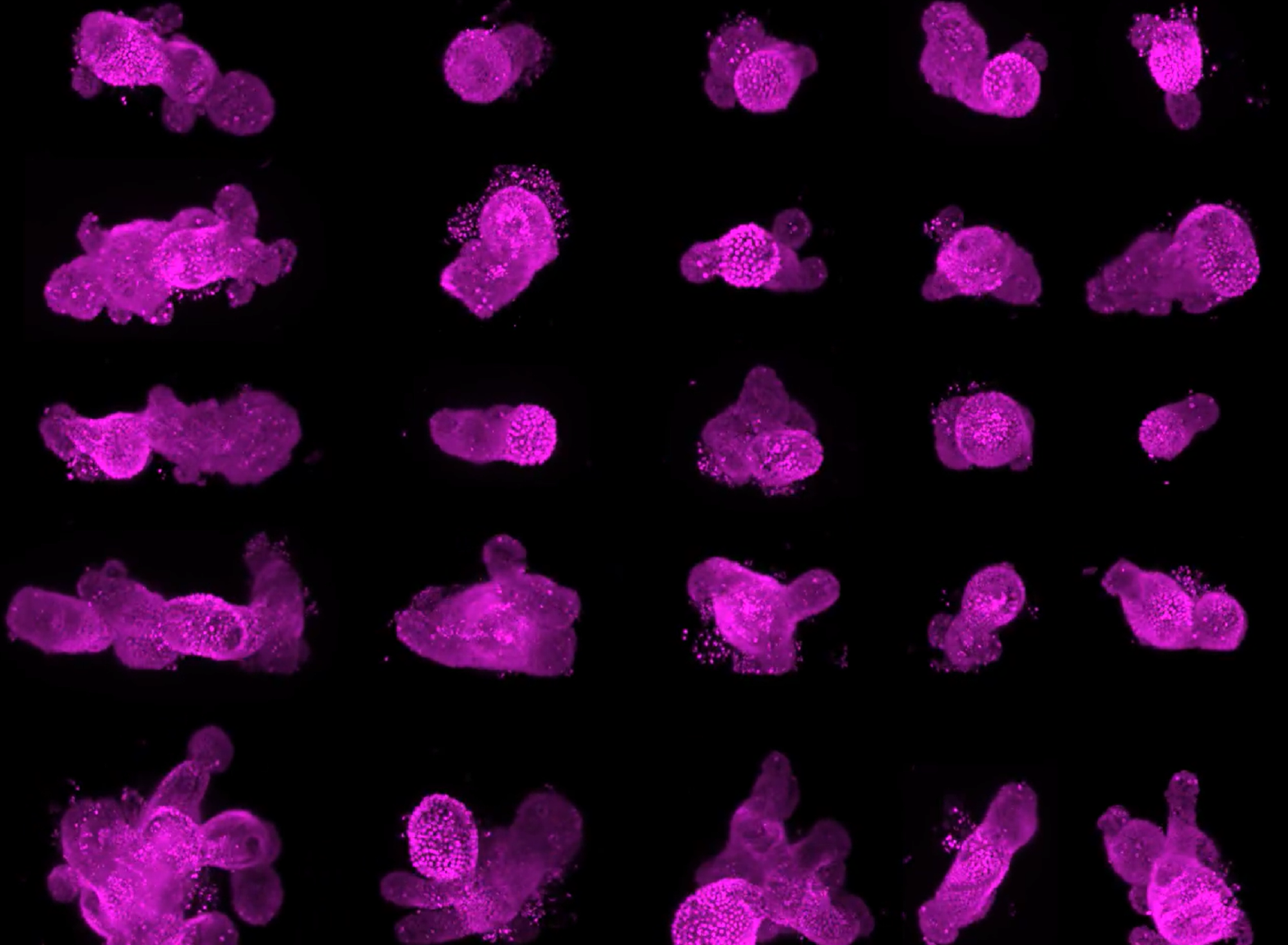
25個成熟的腸道類器官進行了長期多位置成像 (每個類器官的體積為360 µm)
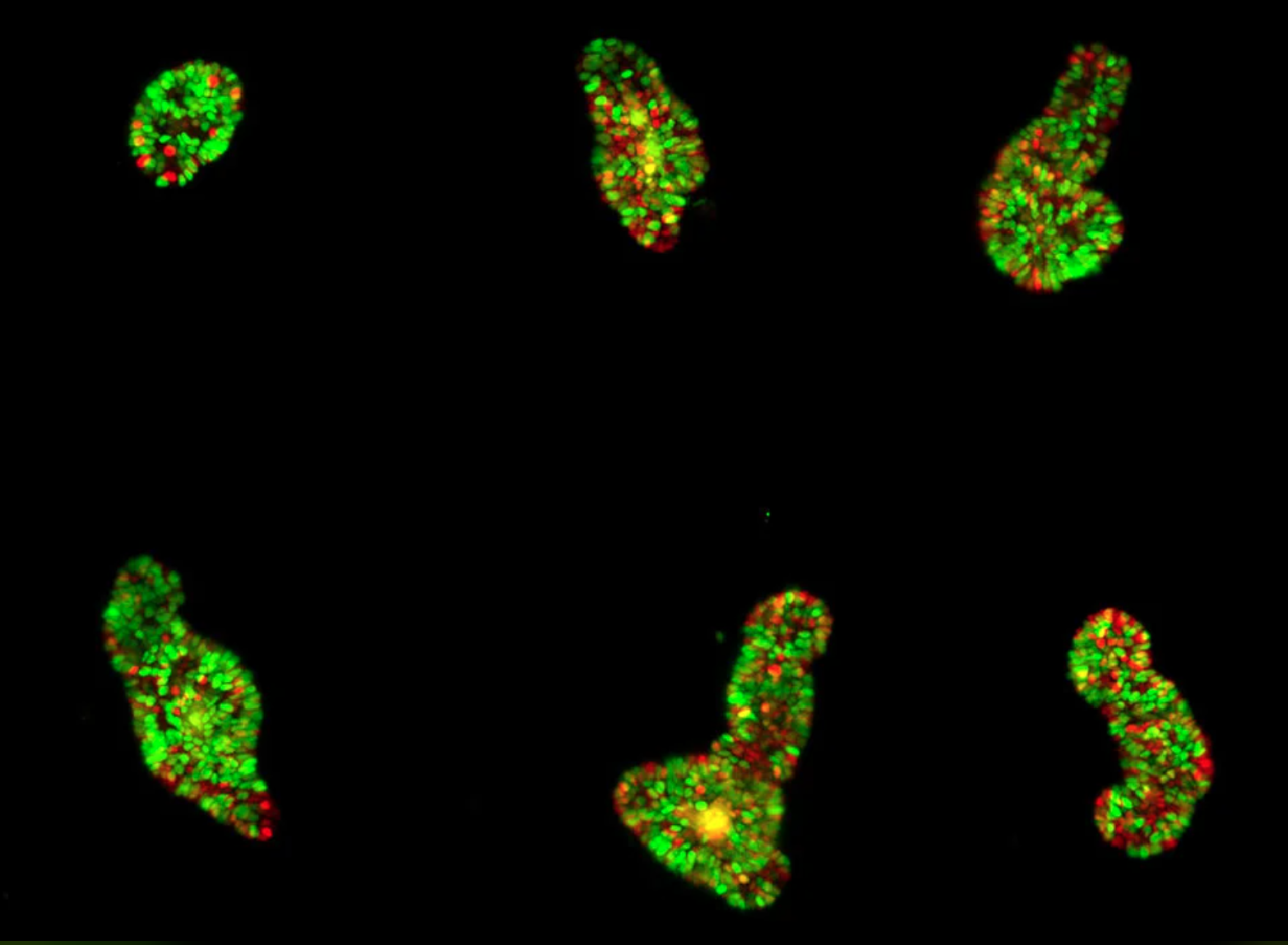 複雜多細胞系統的動態可視化是探索生物生命的一個基本目標。為了解決在大型時空尺度上進行即時成像的挑戰, Viventis Deep採用開放式多樣本雙視角光片顯微鏡。 可以實現單細胞分辨率對大樣本進行成像.
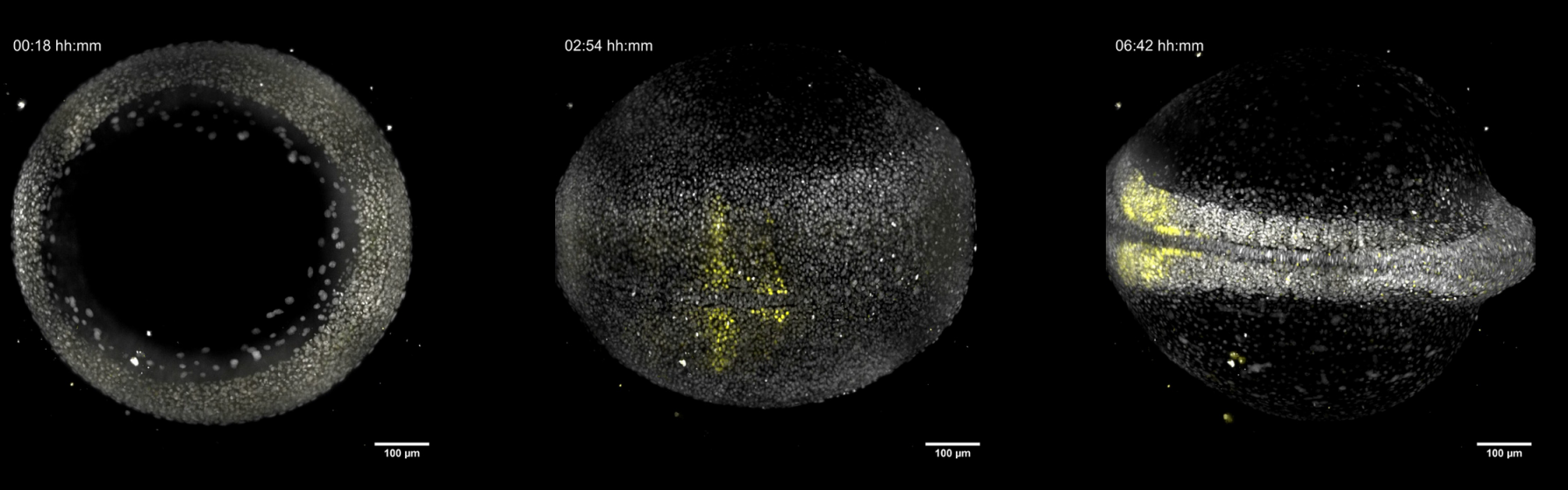 Imaging of cycling gene (yellow) revels timing of somitogenesis in zebrafish embryo. Courtesy of Olivier Venzin, Oates Lab. EPFL Lausanne (Switzerland)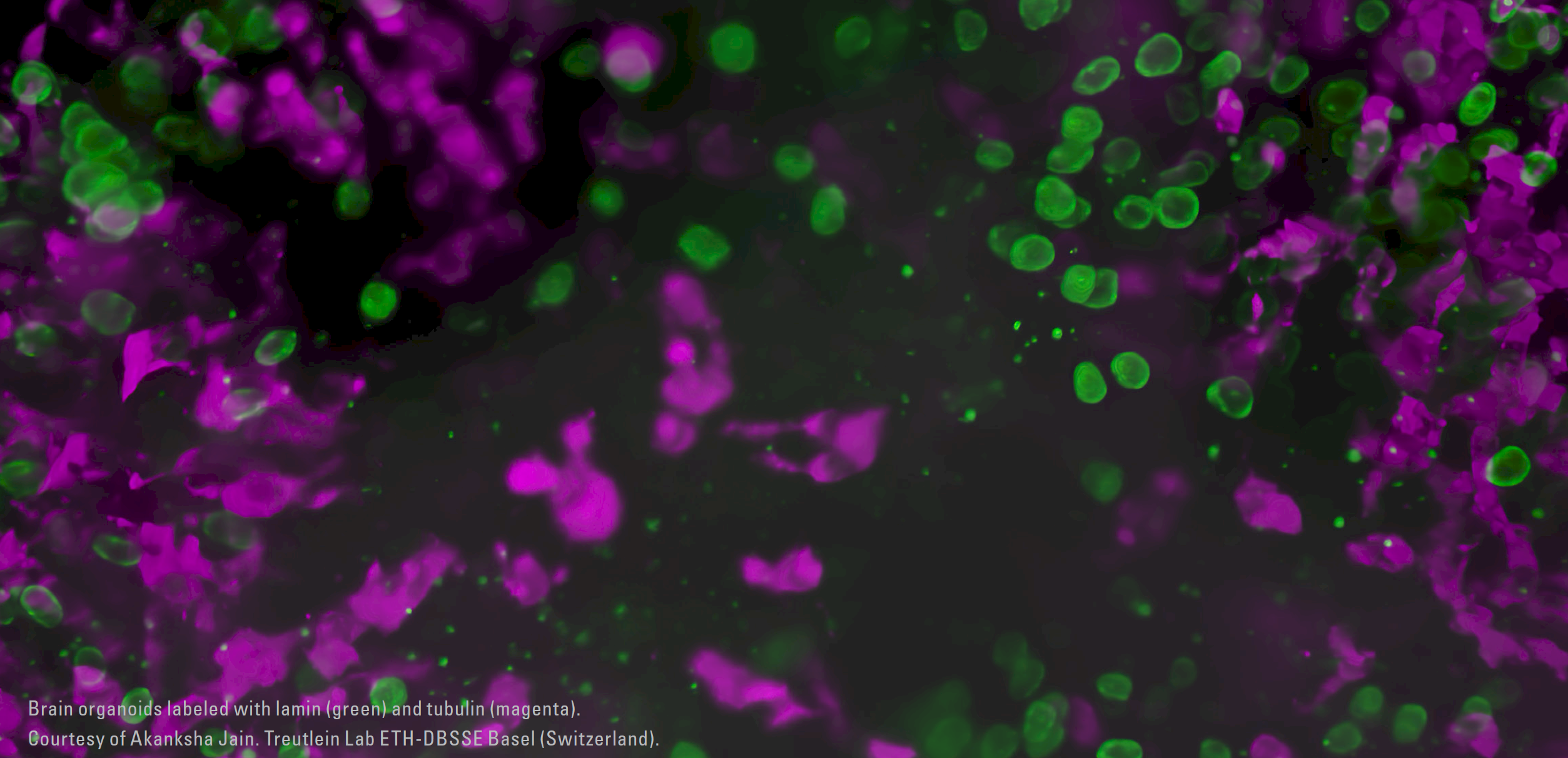
Publications by application
Organoids and 3D cell culture
Tuft cells act as regenerative stem cells in the human intestineHuang et al.
Nature 2024
Mouse neural tube organoids self-organize floorplate through BMP-mediated cluster competition
Krammer et al.
Developmental Cell 2024
NOTCH-driven oscillations control cell fate decisions during intestinal homeostasis
Weterings et al.
BioRxiv (2024). doi.org/10.1101/2024.08.26.609553
Control of lumen geometry and topology by the interplay between pressure and cell proliferation rate in pancreatic organoids
Lee et al.
BioRxiv 2024
The G1/S transition in mammalian stem cells in vivo is autonomously regulated by cell size
Xie et al.
BioRxiv 2024
Open top multi sample dual view light sheet microscope for live imaging of large multicellular systems
Moos et al.
Nature Methods 2024.
Decoding morphogen patterning of human neural organoids with a multiplexed single-cell transcriptomic screen
Sanchís-Calleja et al.
BioRxiv 2024
Integrating single-cell imaging and RNA sequencing datasets links differentiation and morphogenetic dynamics of human pancreatic endocrine progenitors
Beydag-Tasöz et al.
Developmental Cell 2023
Dynamics and plasticity of stem cells in the regenerating human colonic epithelium
Oost et al.
BioRvix 2023
Morphodynamics of human early brain organoid development
Jain et al.
BioRvix 2023
Topological morphogenesis of neuroepithelial organoids
Ishihara et al.
Nature Physics 2022
Multiscale light-sheet organoid imaging framework
de Medeiros et al.
Nature Communication 2022
Cell fate coordinates mechano-osmotic forces in intestinal crypt formation
Yang et al.
Nature Cell Biology 2021
Lineage recording in human cerebral organoids
He
Nature Methods 2021
Tissue Engineering with Mechanically Induced Solid-Fluid Transitions
Mailand et al.
Ad. Materials 2021
Capturing Cardiogenesis in Gastruloids
Rossi et al.
Cell Stem Cell 2021
Self-organization and symmetry breaking in intestinal organoids development
Serra et al.
Nature 2019
Oocytes and Embryos Development
Inverse blebs operate as hydraulic pumps during mouse blastocyst formationSchliffka et al.
Nature Cell Biology 2024
Spatio-temporal requirements of Aurora kinase A in mouse oocytes meiotic spindle building
Blengini et al.
iScience 2024
Live imaging human embryos reveals mitotic errors and lineage specification prior to implantation
Abdelbaki et al.
BioRxiv 2024
Reconstitution of chromatin reorganization during mammalian oocyte development
Wang et al.
BioRxiv 2024
Live-imaging reveals Coordinated Cell Migration and Cardiac Fate Determination 2 during Mammalian Gastrulation
Abukar et al.
BioRxiv 2023
CHK1-CDC25A-CDK1 regulate cell cycle progression and protect genome integrity in early mouse embryos
Knoblochova et al.
Embo Reports 2023
Actin-driven chromosome clustering facilitates fast and complete chromosome capture in mammalian oocytes
Harasimov et al.
Nature Cell Biology 2023
Cell fragmentation in mouse preimplantation embryos induced by ectopic activation of the polar body extrusion pathway
Pelzer et al.
Embo Journal 2023
Mechanism of spindle pole organization and instability in human oocytes
So et al.
Science 2022
Aurora kinase A is essential for meiosis in mouse oocytes
Blengini et al.
Plos Genetics 2021
Hydraulic fracturing and active coarsening position the lumen of the mouse blastocyst
Dumortier et al.
Science 2019
Primed Track, high-fidelity lineage tracing in mouse pre-implantation embryos using primed conversion of photoconvertible proteins
Welling et al.
Elife 2019
Zebrafish Research
LiverZap: a chemoptogenetic tool for global and locally restricted hepatocyte ablation to study cellular behaviours in liver regenerationAmbrosio et al.
Development 2024
Cell-autonomous generation of the wave pattern within the vertebrate segmentation clock
Rohde et al.
eLife 2024
Clock driven waves of Tbx6 expression prefigure somite boundaries
Venzin et al.
BioRxiv 2023
Deconstructing body axis morphogenesis in zebrafish embryos using robot-assisted tissue micromanipulation
Ozelci et al.
Nature Communications 2022
Left-right symmetry of zebrafish embryos requires somite surface tension
Naganathan et al.
Nature 2022
Mitochondria in Embryogenesis: An Organellogenesis Perspective
Arribat et al.
Frontiers in Cell and Developmental Biology 2019
Marine Organisms
Life-cycle-coupled evolution of mitosis in close relatives of animalsShah et al.
Nature 2024
The nuclear-to-cytoplasmic ratio drives cellularization in the close animal relative Sphaeroforma arctica
Marine Olivetta et al.
Current Biology 2023