全部商品 / LEICA 組織切片機與HE組化設備 / Leica HistoCore 病理組織切片機

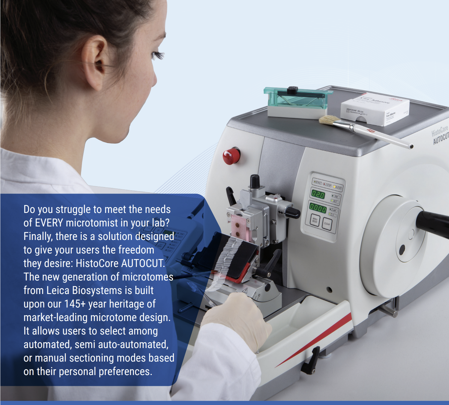
AUTOCUT 病理組織切片機
使用者可根據個人操作偏好, 選擇三種石蠟組織切片模式 :
全自動模式 (一鍵按鈕, 完全自動切片)
半自動 (用按鈕驅動樣品前進和回縮)
手動切片模式
全自動模式 (一鍵按鈕, 完全自動切片)
半自動 (用按鈕驅動樣品前進和回縮)
手動切片模式
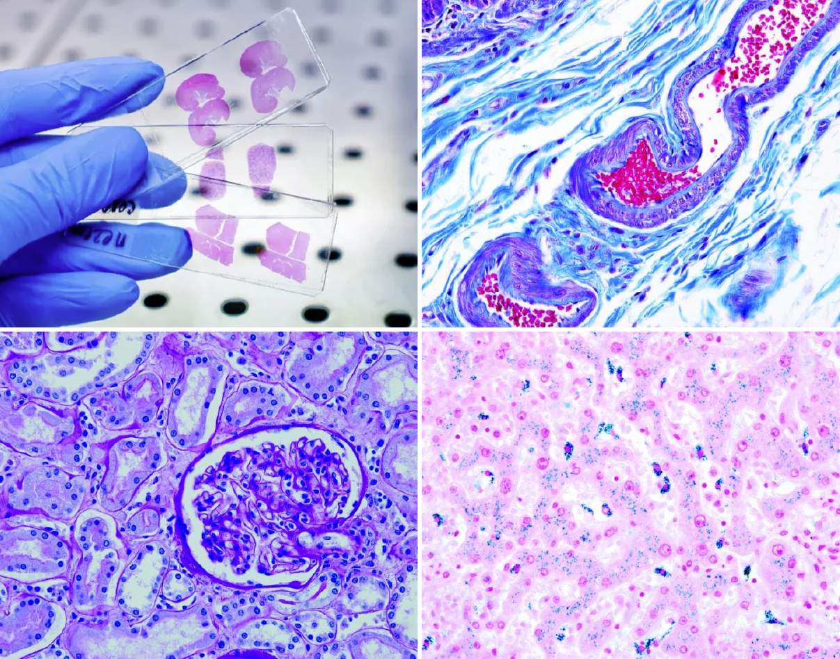
右上圖
Gomori Trichrome (blue) (submucosa). Trichrome stains are used to stain and identify muscle fibers, collagen and nuclei. They can be used to contrast skeletal, cardiac or smooth muscle. The Gomori Trichrome is a simplification of the more elaborate Masson trichrome stain and combines the plasma stain (chromotrope 2R) and connective tissue stain to provide a brilliant contrasting picture.
左下圖
Periodic Acid Schiff (kidney). PAS staining is mainly used for staining structures containing a high proportion of carbohydrates such as glycogen,glycoproteins, proteoglycans typically found in connective tissues, mucus and basement membranes. Often used to stain kidney biopsies, liver biopsies, certain glycogen storage diseases in striated muscles and suspected fungal infections.
右下圖
Perls’ Prussian Blue Iron (liver). This stain is used to detect and identify ferric (Fe3+) iron in tissue preparations, blood smears,or bone marrow smears. Minute amounts of ferric iron (haemosiderin) are commonly found in bone marrow and in the spleen. Abnormal amounts of iron can indicate hemochromatosis and hemosiderosis.
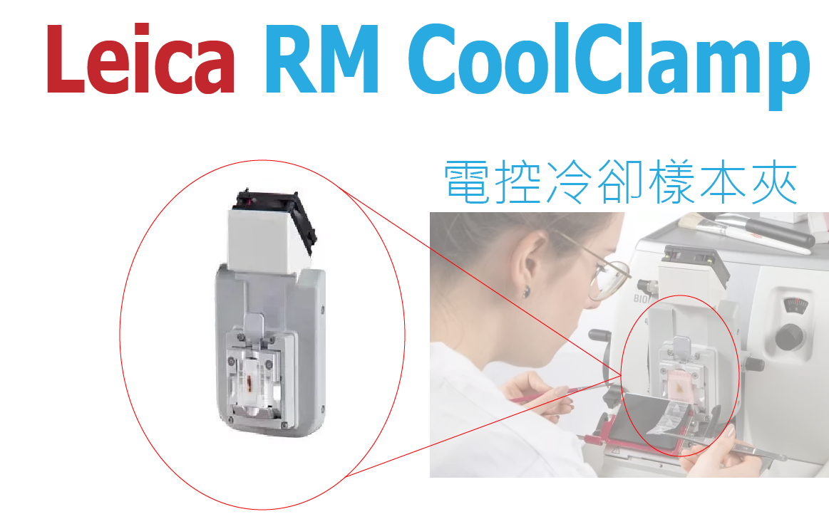
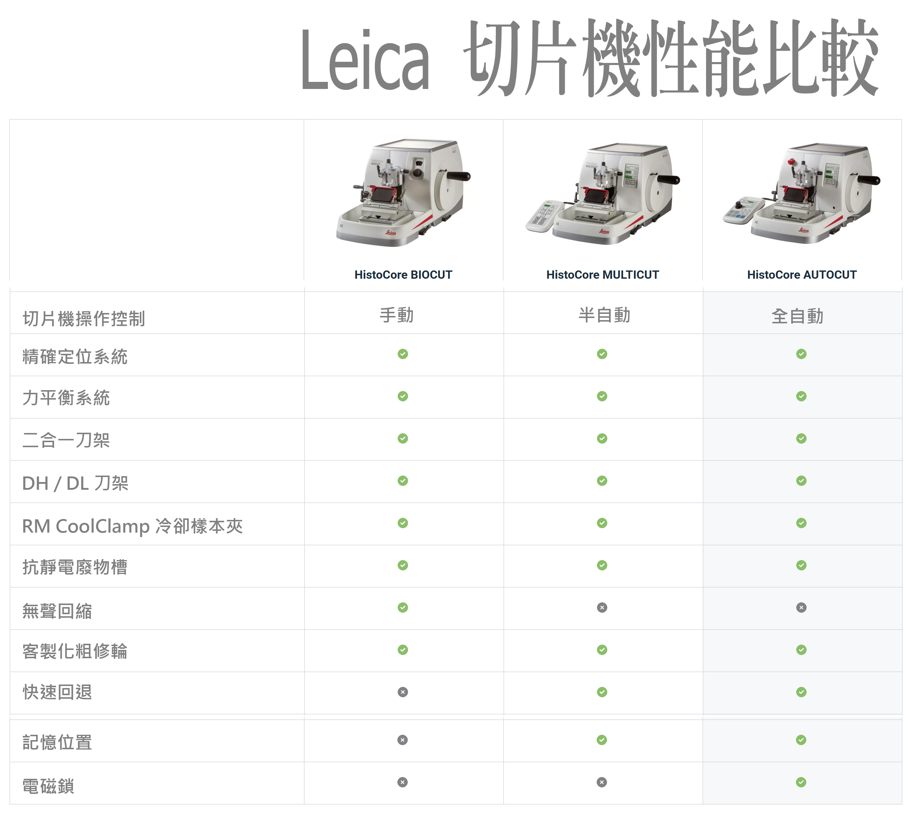
- CoolClamp 電控冷卻樣本夾, 可安裝到任何 Leica HistoCore 系列的切片機 (獨一的設計)
- 將切片溫度保持在環境溫度以下 20°C (無須預先冷卻蠟塊, 在任何環境下, 可隨時切片)
- 可連續切出均勻的切片, 維持最高品質 (每個組織塊都能保持在理想的溫度)
- 輕鬆的使用, 提高切片效率與切片品質 (符合使用者的切片期待)
- 使用於 常規染色、特殊染色和免疫組織化學(IHC)/原位雜交(ISH)染色。
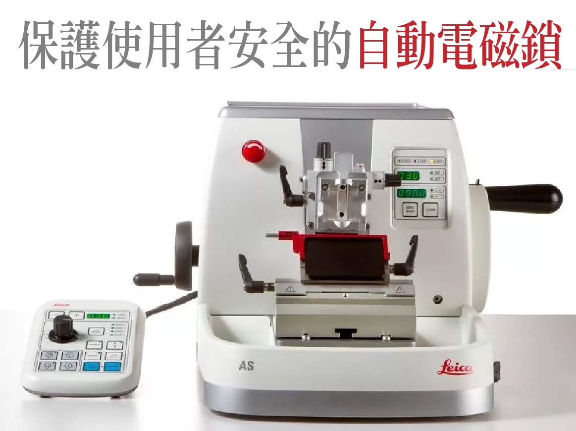 獨家的設計 !
保護使用者安全的自動電磁鎖
暫停自動切片功能時,電磁鎖將立即啟動,將樣品退回並保持在遠離刀片的最安全位置。這可極大降低操作員操作樣品時的受傷風險。
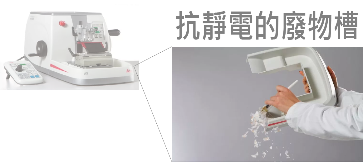
Leica 抗靜電廢物槽, 盡是為使用者設想, 讓切片工作更輕鬆自在. 可保持工作場所清潔、高效且不留碎屑。可防止廢石蠟粘附在槽壁上。其高效無比,可將清潔時間從幾分鐘縮短至幾秒鐘

| 樣品進量: | 約 24 mm ±1 mm |
| 垂直行程: | 70 mm ±1 mm |
| 標準樣品夾:55 x 50 x 30 mm 超大樣品夾:68 x 48 x 15 mm |
|
| 電動切片速度: | 0 - 420 mm/s ±10% |
| 樣品回縮 | |
| 在手動切片模式中: | 5 - 100 µm (5 µm 遞增);可關閉 |
| 在電動切片模式中: | 取決於速度;可關閉 |
| 樣品定位 | |
| 水準: | ± 8° |
| 垂直: | ± 8° |
| 切片厚度設置 | |
| 範圍: | 0.5 - 100 µm |
| 設置值: | 0.5 - 5 µm (0.5 µm 遞增) |
| 5 - 20 µm (1 µm 遞增) | |
| 20 - 60 µm (5 µm 遞增) | |
| 60 - 100 µm (10 µm 遞增) |
| 修片厚度設置: | |
| 範圍: | 1 - 600 µm |
| 設置值: | 1 - 10 µm (1 µm 遞增) |
| 10 - 20 µm (2 µm 遞增) | |
| 20 - 50 µm (5 µm 遞增) | |
| 50 - 100 µm (10 µm 遞增) | |
| 100 - 600 µm (50 µm 遞增) |
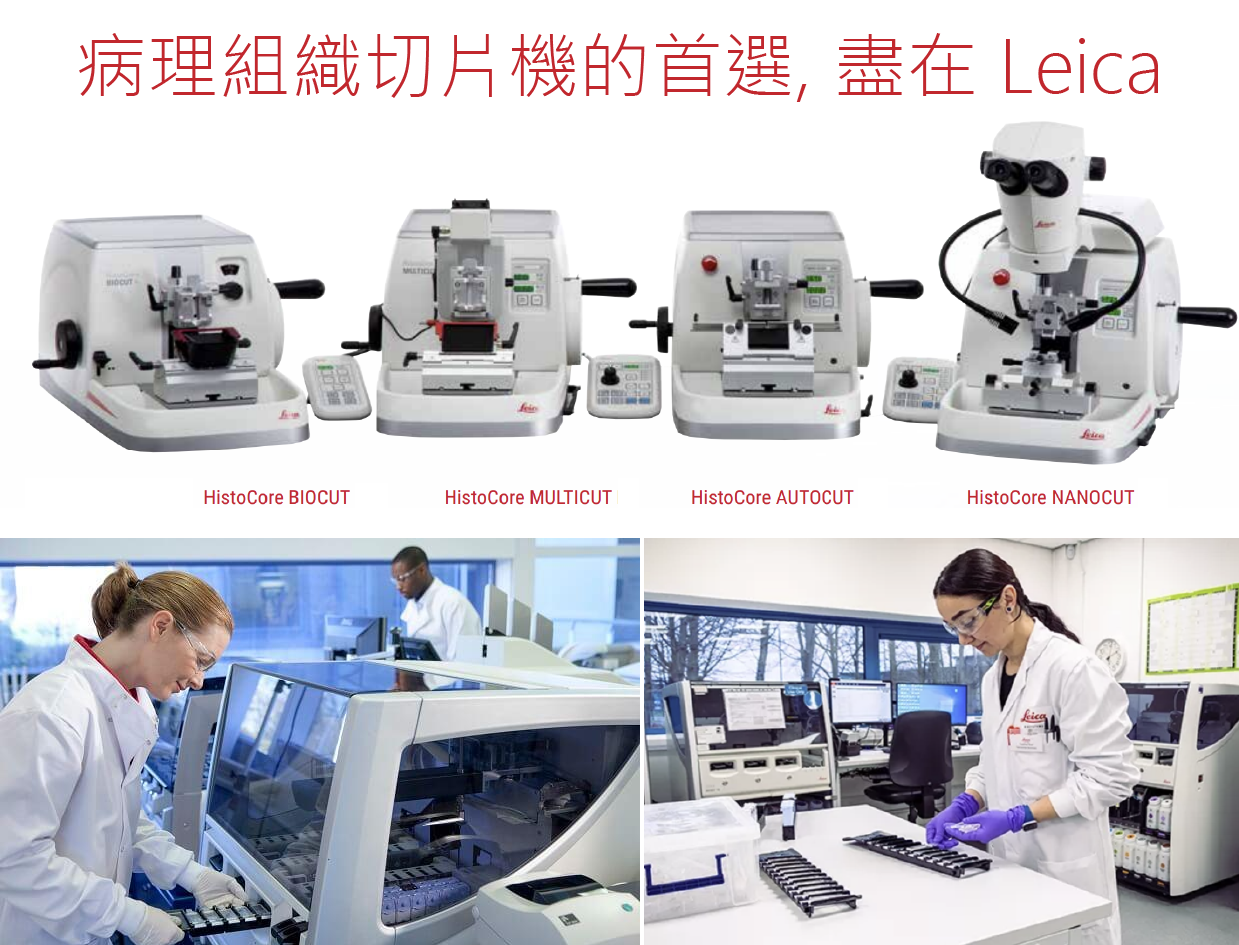
AUTOCUT 提供優質的切片, 提升診斷的精準
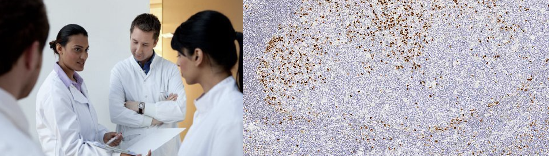 多發性骨髓瘤致癌基因1(MUM1)
MUM-1(多發性骨髓瘤癌基因1)基因最初被發現是因為它參與了多發性骨髓瘤中觀察到的t(6:14)易位,導致了MUM-1基因與Ig重鏈位點並置。MUM-1在B細胞分化的漿細胞定向晚期和活化的T細胞中均有表達,提示MUM-1可作為這些細胞衍生的淋巴造血腫瘤的標誌物。已發現表達MUM-1的細胞的形態學光譜範圍從中心體細胞到漿母細胞/漿細胞。因此,MUM-1的組織發生價值可能為鑒定BCL-6陽性(生殖中心B細胞)向CD138陽性(免疫母細胞和漿細胞)轉化提供了一個標記。