全部商品 / LEICA 正立光學顯微鏡 / LEICA LMD 雷射顯微組織切割系統

全自動雷射顯微單細胞與組織掃描切割擷取系統
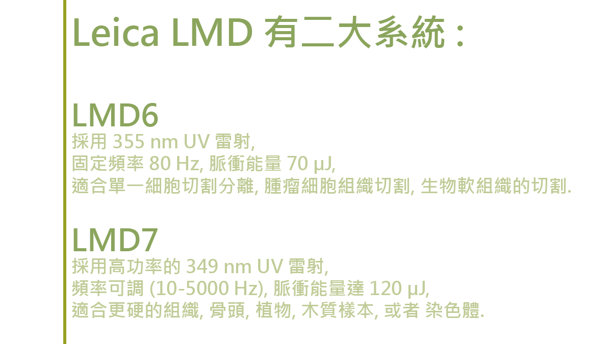
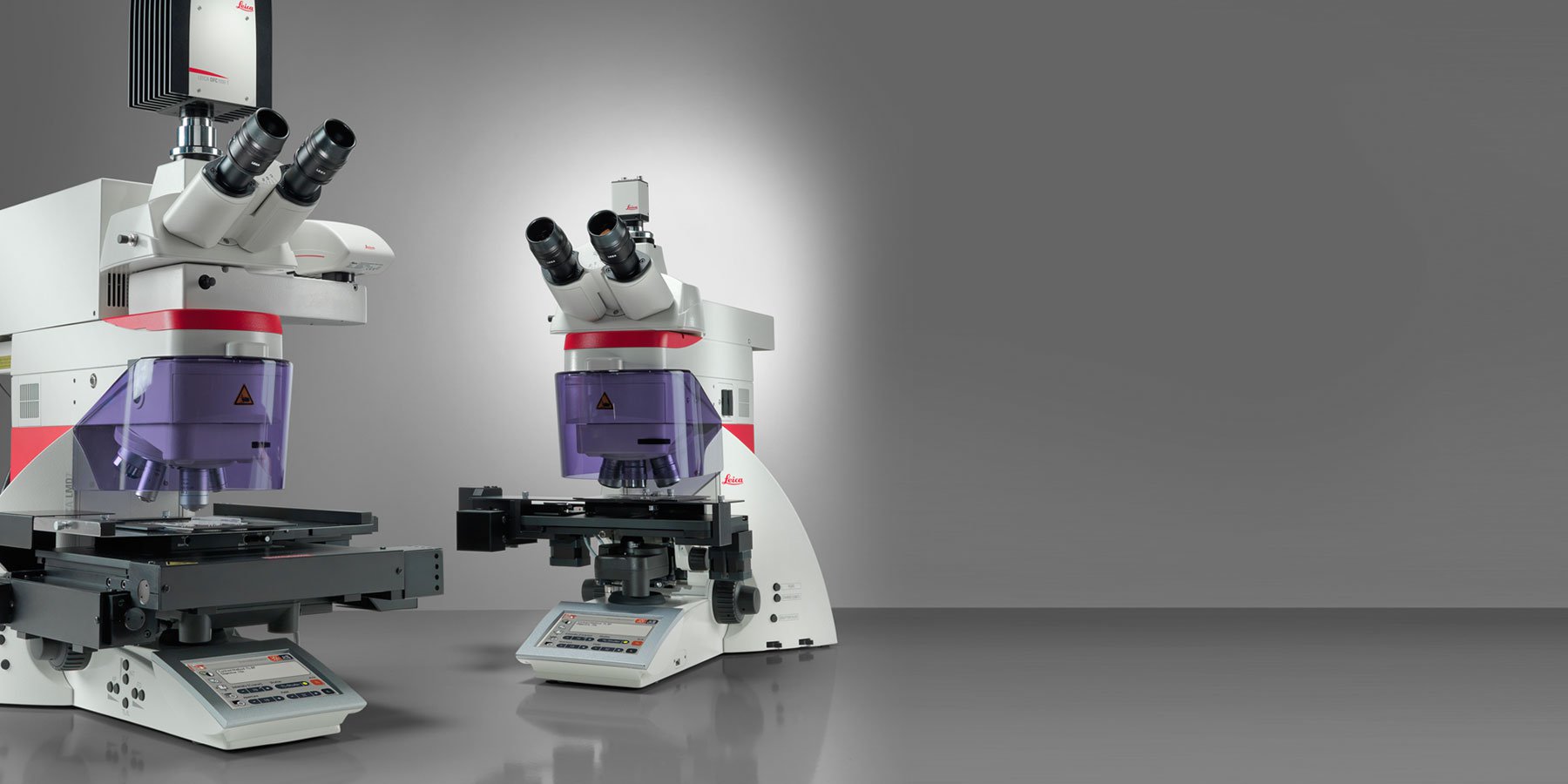
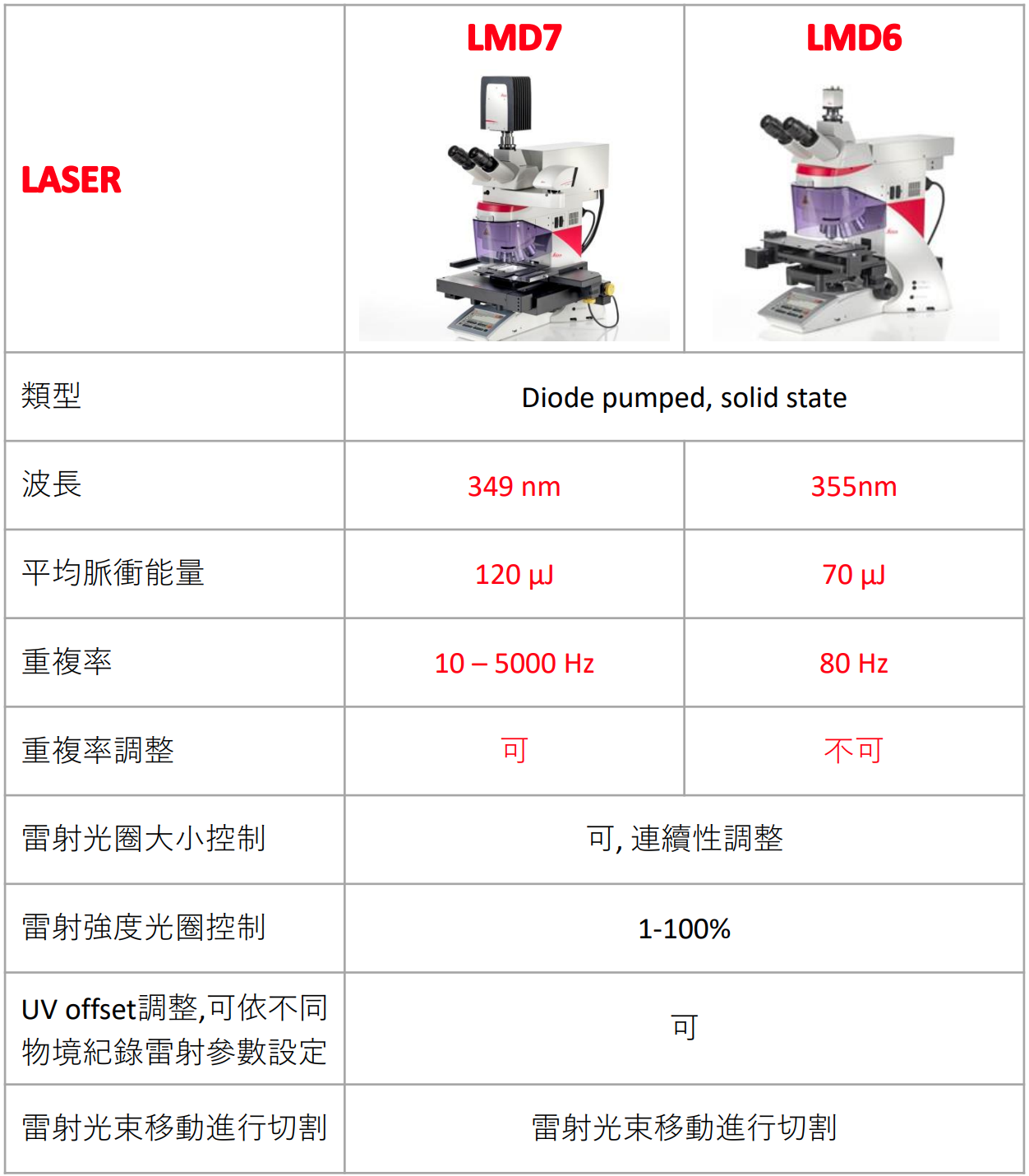
採用 355 nm UV 雷射, 80 Hz, 脈衝能量 70 µJ, 適合單一細胞切割分離, 腫瘤細胞組織切割, 生物軟組織的切割.
LMD7 :
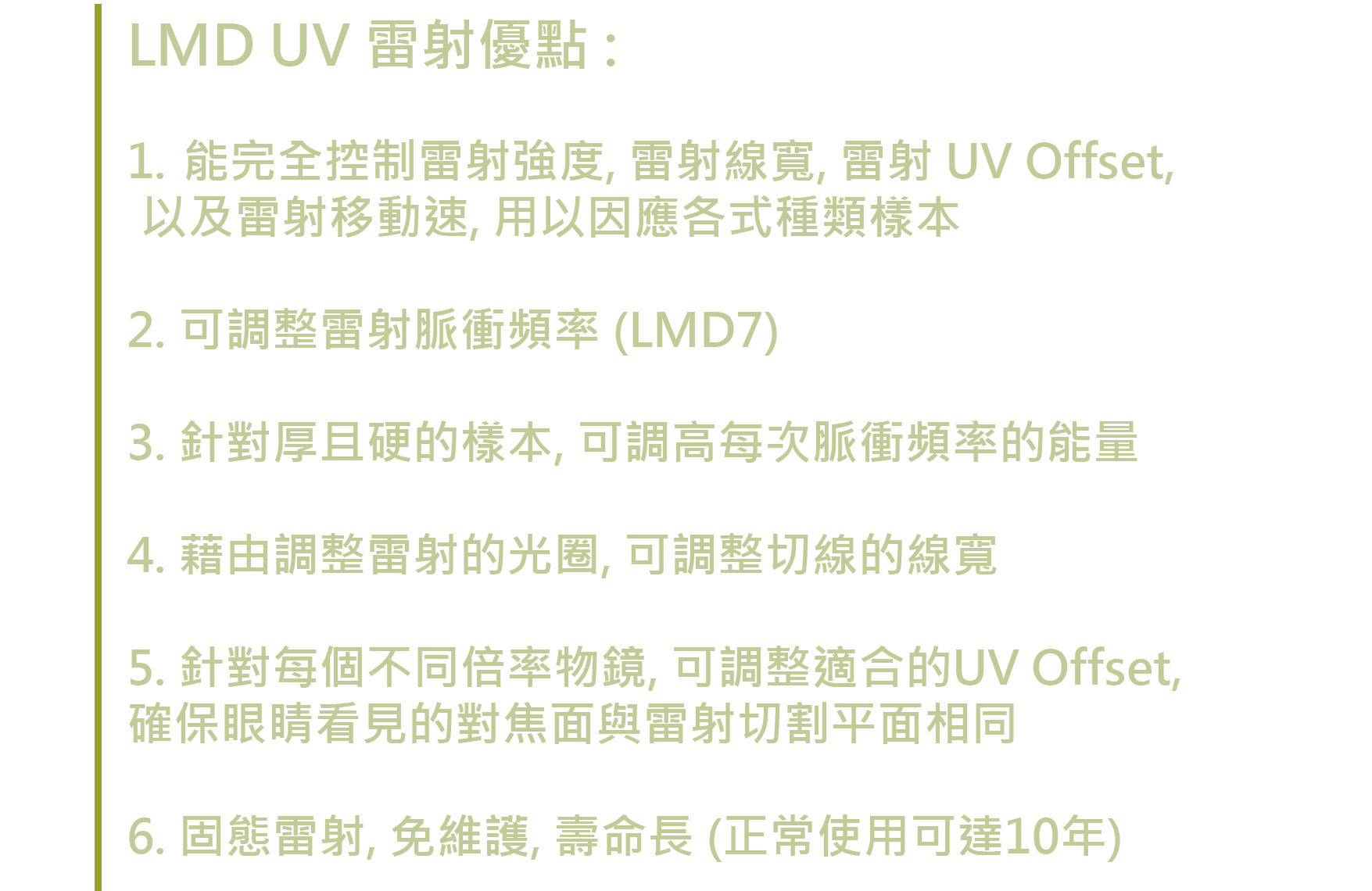
採用高功率的 349 nm UV 雷射, 頻率可調 (10-5000 Hz), 脈衝能量達 120 µJ, 適合更硬的組織, 骨頭, 植物, 木質樣本, 或者 染色體.


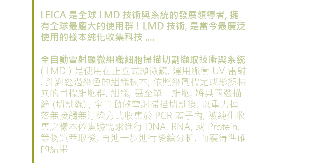


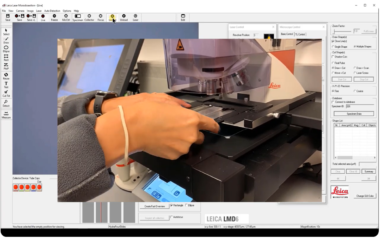
以下影片, 介紹 LMD 的使用過程 ...

Leica LMD 是該 " 顯微單細胞與組織切割擷取 " 的領航者, 具有全球公認的重大優勢 :
1. 在正立式顯微鏡下, 使用脈衝 UV 雷射掃描切割 (可針對樣本特性, 做雷射的最佳調整設定), 同時具備精準與高速切割的兩大效益.
2. 使用者可任意圈選擬切割的組織樣本區域, 即可點擊全自動掃描切割, 使用上的簡易便捷, 省時省力, 輕鬆地提高工作效率. (可以大量精準取得最純的樣本) - 獨家使用 ADM 人工智能辨識樣本, 可以通過 AIVIA 辨識複雜的樣本, 快速自動圈選並切割所要的樣本物件, 導入 LMD 切割, 在短時間內收集大量樣本, 達到智能自動化的切割取樣 !!!
3. 被切割後的樣本, 以重力掉落方式收集於 PCR 蓋子內, 完全做到無接觸無汙染, 達到純化取樣的終極目標. 對後續的分析, 有重大的意義.
4. 整個切割取樣過程, 完全可視化, 可錄影拍照存查. 確保品質可靠性.
5. 可應用到單一細胞取樣, 活細胞取樣.
6. 具有獨一的 LMD 專屬切割物鏡 ( 2.5x – 150x ).
7. 可應用於螢光切割取樣.
8. 豐富的各式樣本耗材以及各式收集方式, 用以提高應用範疇.
LMD 主要應用
• Cancer research
• Pathology
• Molecular biology research
• Neuroscience research
• Developmental research
• Plant research
• Forensics
• Physiology research
• Clinical research
• Pharmaceutical research
除上述應用, LMD 的雷射系統也可在微小個體如 : Drosophila, C. elegant, 或 Zebra Fish…等樣本, 進行所謂的微型手術 (Micro-Surgery), 進而觀察手術後之變化

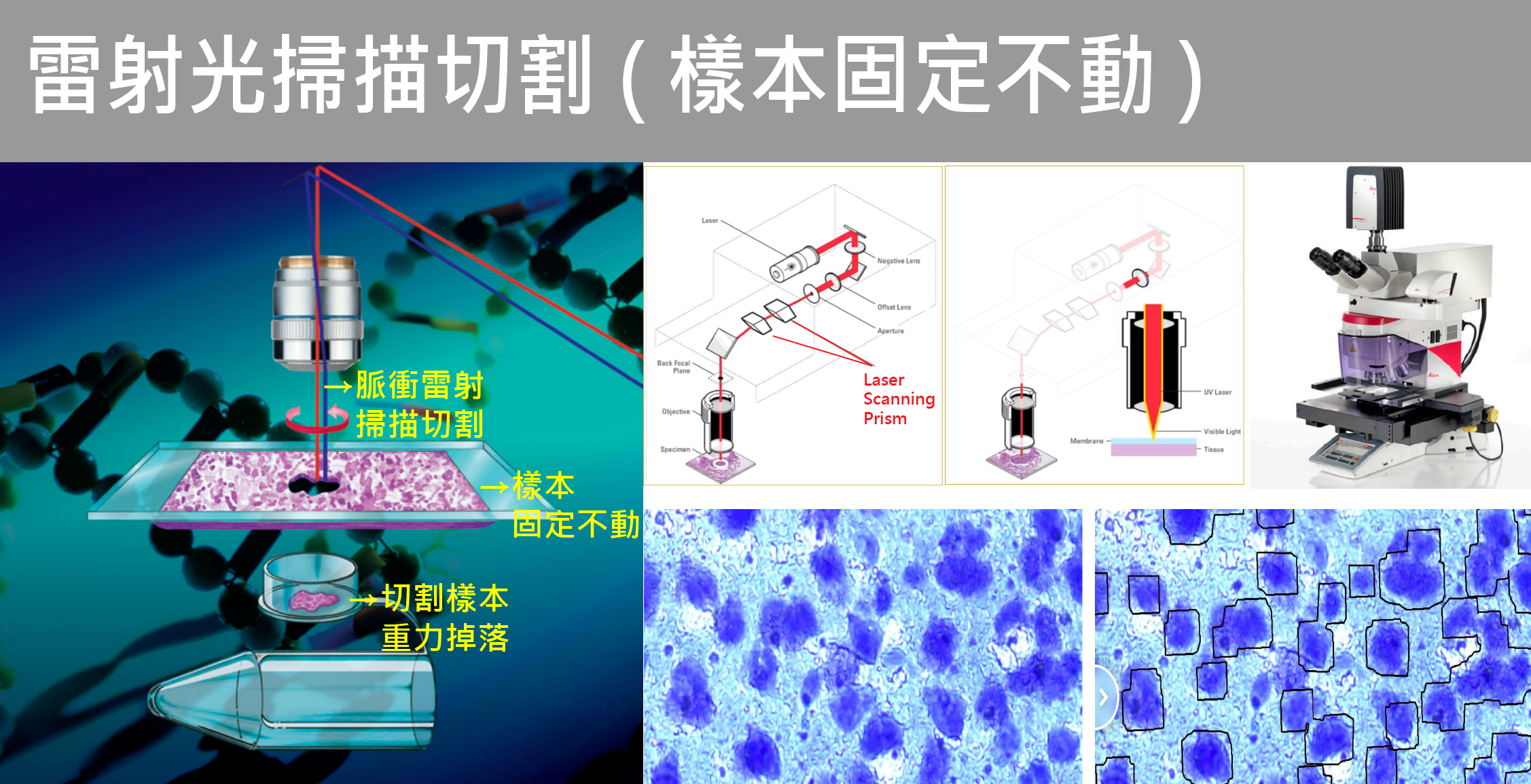
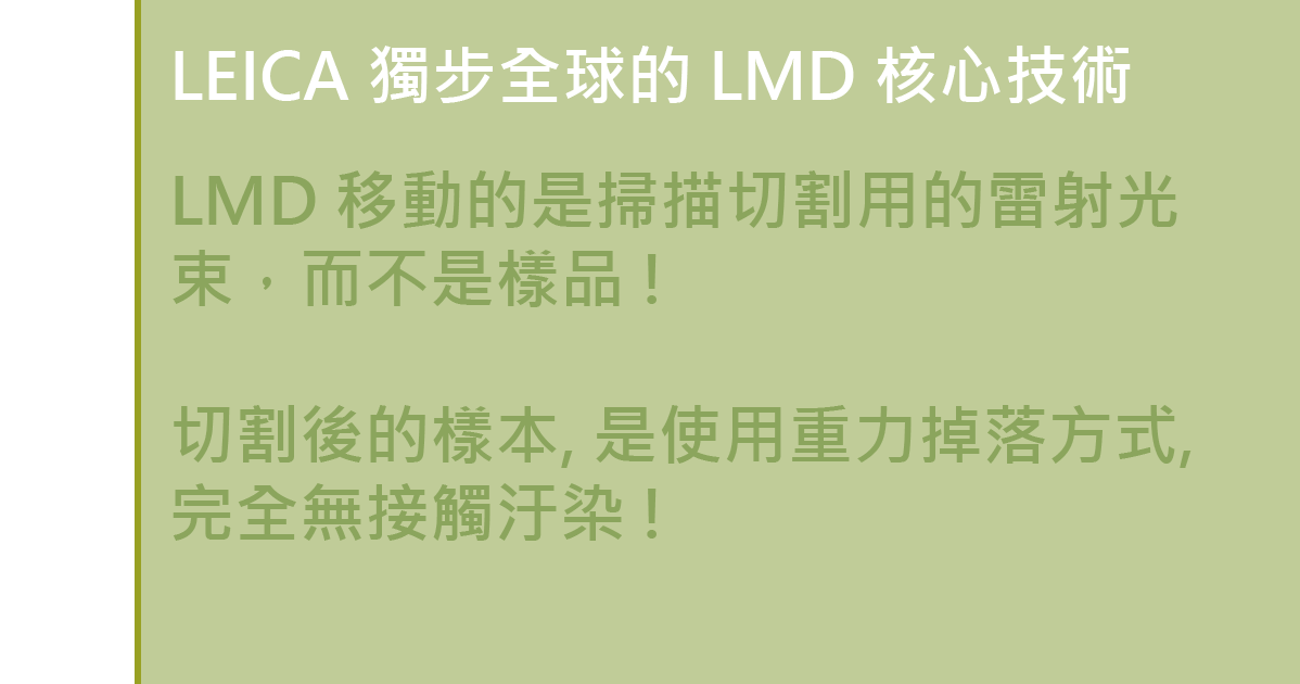
我們移動的是切割用的紫外雷射光束,而不是樣品 !
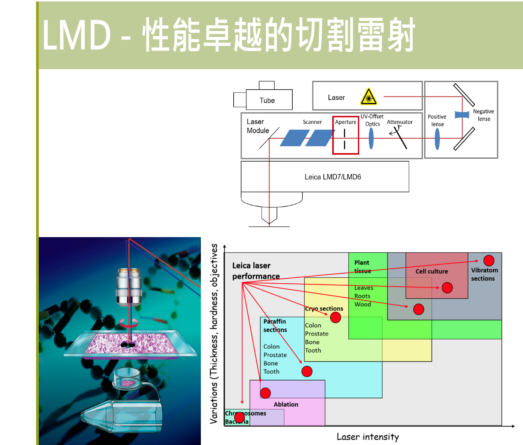
這是 LEICA LMD 獨創的先進顯微切割技術, 通過全自動化的掃描棱鏡, 精準的沿著組織樣本上的指定切割線, 雷射光束會精準的自動掃描切割, 所創造出獨一的優勢 :
1. 最精準的顯微樣本切割, 始終如一 !
2. 高速掃描, 高速切割 ! 最高效率 !
3. 使用簡單與快速, 可以切割任何形態複雜的樣本, 可以大量快速切個樣本 !
4. 雷射光徑與強度皆可調 ( Aperture / Offset ) , 可以切割更小或更厚的樣本, 迎刃而解 !
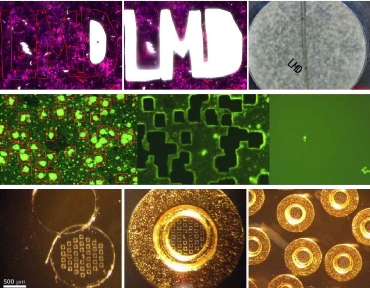

對於形態複雜的大量樣本區, 又計畫大量切割取樣, Leica LMD 又一個人工智能得自動樣本型態偵測軟體 ( ADM ), 可以省時省力的瞬間取得切割樣本, 一鍵輕易切割取樣.

The ADM (Auto Detection Mode) is a software module which helps collect large amounts of similar dissectates. You just have to encircle a region you are interested in and the software will use this as a template for other regions. For example, Proteomics analysis requires a large amount of sample to be captured, and ADM has provided a great benefit to labs working with 100's-1000's of dissectates.
Autofocus can be used before each dissection process to keep the focus of hundreds of ROIs distributed over the slide. Moreover, you can automatically inspect and document all collection devices which were used.
The systematic approach: A raster tool separates your Field of View into a given number of regions. With this function, you can systematically dissect your specimen and collect your dissectate into a variety of collection devices such as 96-Well plates.

獨一 的 ADM 優點 : ( 選配軟體 )
• 可應用於穿透光模式以及螢光模式下
• 可依需求選擇灰階強度模式, 或者依顏色篩選出所需物件
• 參數設定可使用手動, 半自動, 以及自動模式, 用 以因應各式樣本
• 篩選出之物件, 仍可進行手動增加或刪除進行修改
• 設定完成之參數可進行儲存, 於下次需使用時進行套用
• 適合並支援高通量 (HTS) high‐throughput support 收集


LMD 顯微切割操控軟體 - 優點 :
• 直觀的圖示化介面, 即使未使用過, 也容易學習快速上手 !
• 提供樣本 Overview 功能, 用以快速找到要切割的區域 !
• 所獲得的 Overview 影像, 可當作導航影像, 於 Overview影像上點選所需位置, 則此位置會移至視野下
• 完全控制顯微鏡, 載物台, 雷射各式參數
• 具有錄影功能, 可錄製切割過程
• 可加購 樣本自動辨識 ADM (AVC) 功能, 用以大量快速收集樣本
• 可加購 Data Base ( LIF-Browser) 功能, 則可自動儲存切割前, 切割後影像, 所儲存的檔案格式.lif檔 也可使用LAS X開啟
AIVIA - 應用於 LMD 的人工智能軟體
- Mund et al., Nature Biotechnology, 2022 https://doi.org/10.1038/s41587-022-01302-5
- Mitchell et al., JoVE, 2022 https://doi.org/10.3791/64171
AIVIA Interface for Artificial Intelligence ( option )
Aivia is our AI-based, image visualization, analysis and interpretation platform. With the help of AI-enhanced tools such as the Pixel Classifier, Aivia can be used to automatically define Regions of interest (ROI) which are destined for Laser Microdissection (LMD). ROIs can be detected by Aivia and imported directly into the LMD software for Microdissection. Besides Aivia other external software can be used for automatic ROI detection. The LMD software simply needs an XML file containing the ROI information.

快速 ! 效率 !
三步驟即完成切割 :
1. 圈選位置
2. 進行切割
3. 重力方式掉 落收集樣本
優點 :
• 在收集樣本過程中無外力介入
• 無接觸, 無汙染
• 快速收集
• 於收集器中直接加入培養液或反應溶液, 則被切割樣本可 直接掉落於液體中
• 可收集大量樣本( 不會受到收集器的限制)
• 可設定不同類型樣本掉落至不同的收集器位置
• 提供各式樣本holder以及各式收集器, 用以因應各式實驗
• 各式收集器,如: 0.2/0.5 PCR tubes, 8 strips…等, 皆可使用實 驗室現有的, 無需特別購買 (其他廠牌使用倒立式顯微鏡, 收集器位於樣本上方, 為了能 收集樣本, 於收集器上蓋需有黏著劑才能將切割樣本黏住)

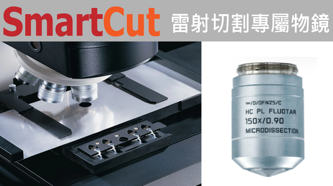

只有 Leica 提供特殊的 LMD 雷射切割專屬物鏡, 優越的性能, 可以提供靈活的紫外光雷射的最佳顯微切割光束設定, 從 2.5x 到 150x ( dry ) 的物鏡, 都具有最高的紫外光 ( UV ) 雷射光傳輸效率,可切割組織、骨骼、牙齒、大腦、植物、染色體和活細胞 – 滿足應用於各類顯微切割的應用!
SmartCut 核心的 LMD 150x 切割物鏡, 可以提供最高的倍率, 查看樣本的細節, 精準執行最精細的切割 ! SmartCut 2.5x 的物鏡, 可以提供最大的視野, 完成最大量與快速的切割.
LMD 切割物鏡具有極高的 UV 穿透率, 具備高效的切割光束外, 也充分利用鏡頭的 NA 值, 提供最高解析的成像效果 !
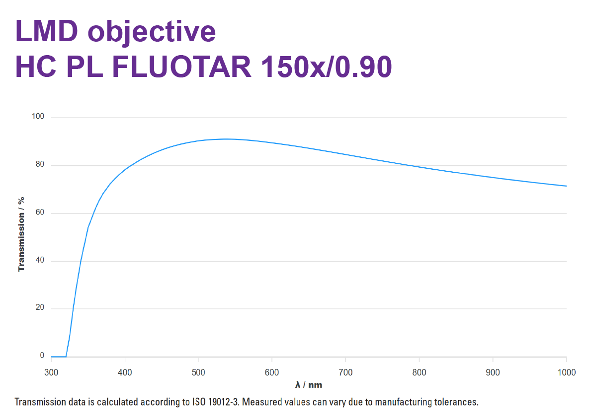
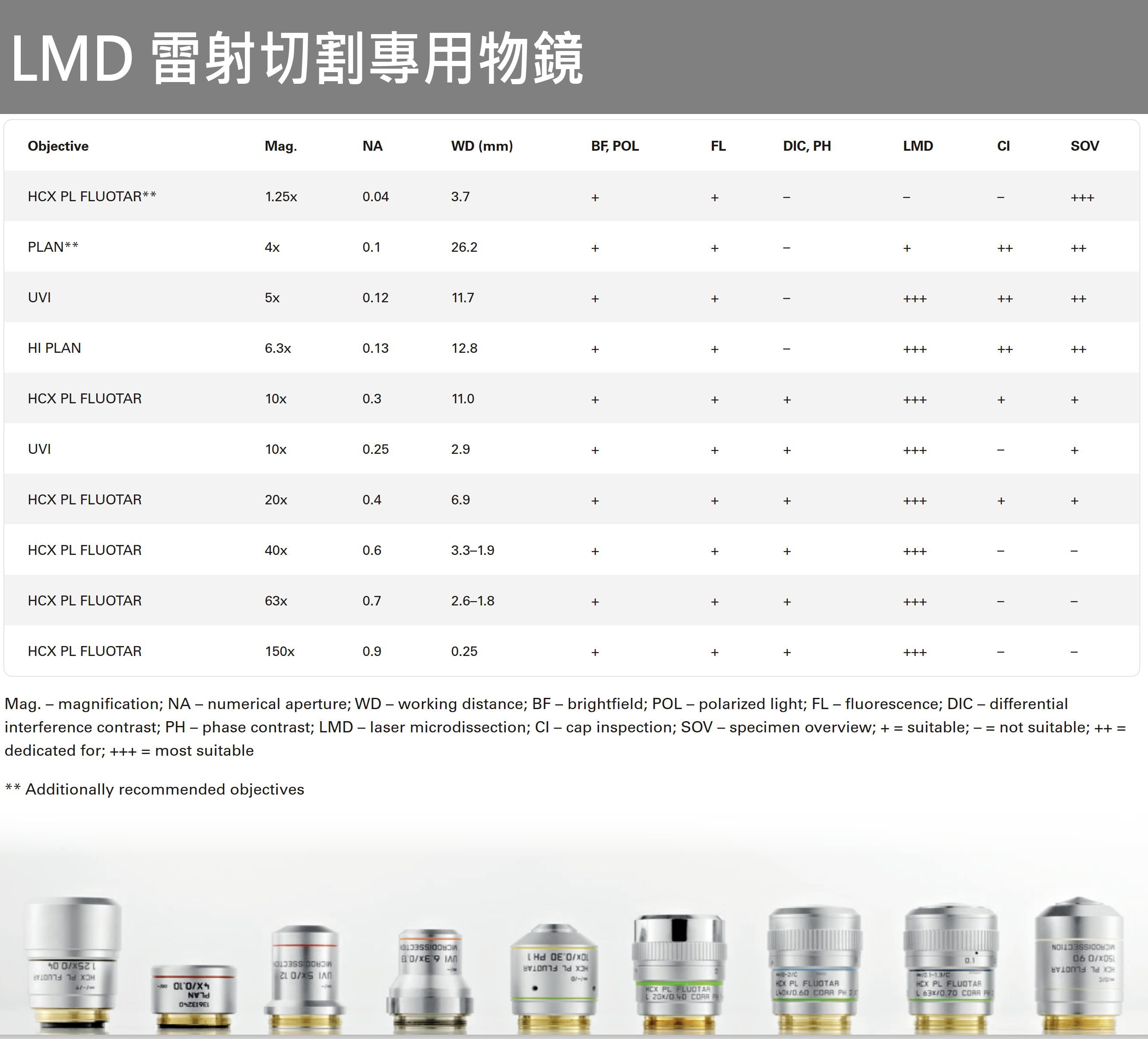
- 提供獨家最高倍 150X Smartcut LMD 專用鏡頭, 用以更精確 切割微小樣本, 如: 染色體 chromosome…等 •
- 2.5X, 5X, 6,3X, 10X, 20X 物鏡適合用於大範圍區域切割 •
- 40X, 63X 物鏡適合用於單細胞切割 •
- 20X, 40X, 63X 物鏡適合應用於 AVC 物件自動辨識功能 •
- 使用 10X 物鏡切割厚度厚達 400 µm 之樣本.- 另有1.25X 鏡頭用於快速樣本的檢視


LMD 螢光樣本切割
提供 LMD 專用螢光濾鏡組進行螢光樣本切割

優點 :
• Leica LMD 為雷射光路與螢光光路各自獨立的系統, 是唯一 可同時觀察到螢光影像與切割過程的系統 (其餘廠牌雷射 光路與螢光光路共用)
• 提供LMD 專用螢光濾鏡組 (於UV波段提高穿透率), 能更有 效率將樣本切割下來
• 提供不同Channel影像疊加功能, 用以找出兩個染色以上皆 表現之物件


Combination System: LMD System with THUNDER Imaging
The LMD6 and LMD7 systems can be combined with THUNDER. The base stand of the THUNDER Imager 3D Tissue and LMD systems are the same, thus such a combination offers:
Lab space savings with one system for different tasks
Laser Microdissection with the LMD without limitation
Brilliant THUNDER fluorescence imaging using LAS X, even in multi-color and 3D (z-stacks) and visualization in the 3D viewer.

Leica LMD 技術原理介紹 ( 影片 )
Laser Microdissection (LMD) is a technique for isolating specific and pure targets from microscopic heterogeneous samples for downstream analysis (DNA, RNA & proteins); based on microscopic imaging and utilizing a laser. In contrast to other systems which use a fixed laserfocus for dissection, Leica Microsystems' LMD systems guide the laserfocus for dissection. This unique feature allows highly precise laser dissection independent of the stage accuracy.
Leica LMD 操作詳細介紹 ( 影片 )
Basic Operation for Cutting & Collecting from Membrane Slides
Leica LMD 6/7: Basic Operation for Cutting & Collecting from Membrane Slides - YouTube
